The ARI laboratory is located at Q150 and is equipped with the following instruments:
- FTIR Microspectroscopy
-
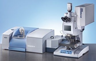
FTIR Microspectroscopy (INVENIO FTIR Spectrometer equipped with HYPERION 3000 Vis-IR Microscope, Bruker Optics) 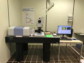
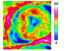
FTIR Microspectroscopy is a powerful and label-free vibrational technique, used to study the chemical composition of biological samples (such as tissues, biological fluids, and cells) and organic and inorganic compounds (including polymers and biocomposites), with a spatial resolution up to ~2.56 mm. FTIR spectroscopy is sensitive to polar (usually antisymmetric) group vibrations. The coupling with the optical microscope allows to correlate the morphological features of biological non-homogeneous samples (such as tissues and cells) with the chemical composition of the sample itself. 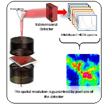
Hyperspectral characterization of the MSTO-211H cell spheroid model: A FPA–FTIR imaging approach
10.1016/j.clispe.2021.100011
The Invenio FTIR spectrometer is coupled with a Hyperion 3000 Vis-IR microscope. It is equipped with a HgCdTe (MCT_A) detector for single point analysis (ca- 20 micron spatial resolution) and a bidimensional Focal Plane Array (FPA) detector for imaging analysis (64×64 pixel/spectra, covering an area of ~164×164 μm2, with a spatial resolution of 2.56 micron). The acquisition of spectra can be performed in reflection or transmission mode in the MIR (Medium InfraRed) region (4000–800 cm−1). 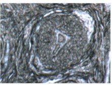
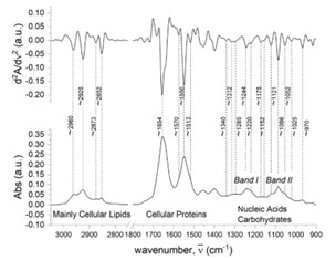
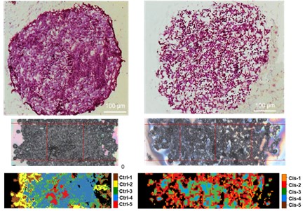
FPA-FTIR analysis of triple-negative breast cancer samples The instrument is also implemented with the Platinum ATR, a single reflection diamond ATR (Attenuated Total Reflectance) accessory, equipped with a DTGS detector, and which can be mounted in the sample compartment of the Invenio interferometer. ATR-FTIR spectroscopy is an in situ technique applied to investigate molecular compounds and biological samples in aqueous systems, enhancing the achievable spatial resolution. 

- Raman Microspectroscopy
-
- Raman Microspectroscopy (XploRA Nano spectrometer, Horiba Scientific)
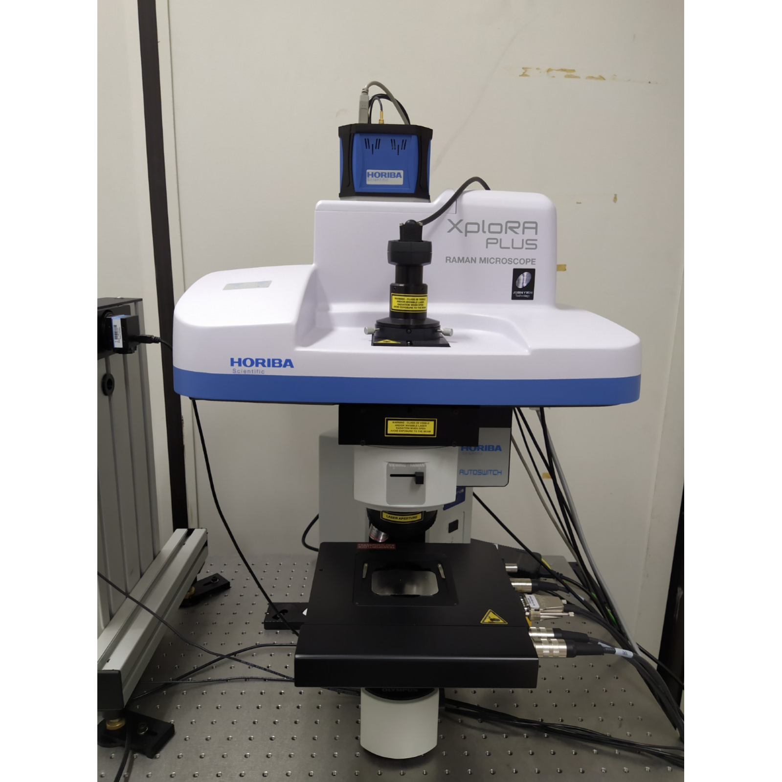
Raman Microspectroscopy is a powerful and label-free vibrational technique, used to study organic, inorganic and biological samples, with a spatial resolution up to ~1 mm. Raman spectroscopy is sensitive to polarizable (usually symmetric) group vibrations. 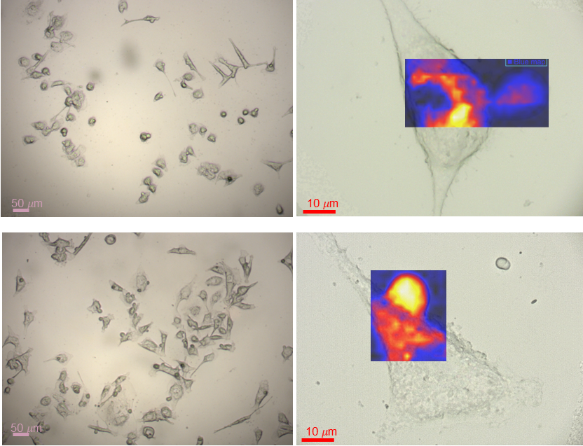
In vitro label-free screening of furanodiene delivery by monoolein cubosomes and cellular effects
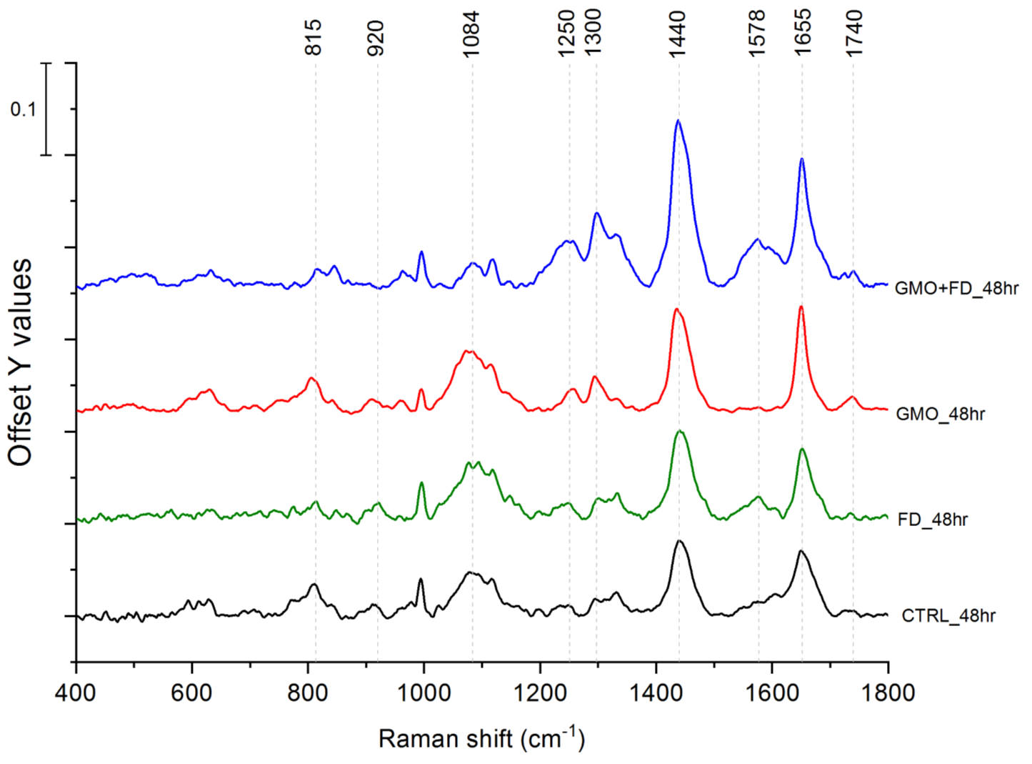
10.1016/j.saa.2021.120735The XploRA Raman microspectrometer is equipped with 532-nm and 785-nm diode lasers. The investigation of the sample with visible light, together with the Raman measurements, can be performed with ×5, ×10 and ×100 objectives (Olympus, N.A. 1). The spectrometer is calibrated to the 520.7 cm−1 line of silicon prior to spectral acquisition. The spectral, spatial and axial resolutions can be set by the regulation of the hole and the slit, and by the selection of the grating (600, 1200, 1800, and 2400 lines per mm). Spectra are dispersed onto a 16-bit dynamic range Peltier cooled CCD detector.

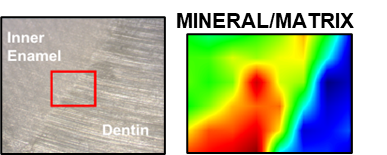
Raman MicroSpectroscopy study of the microstructure and chemical composition of vestibular and lingual surfaces in permanent and deciduous human teeth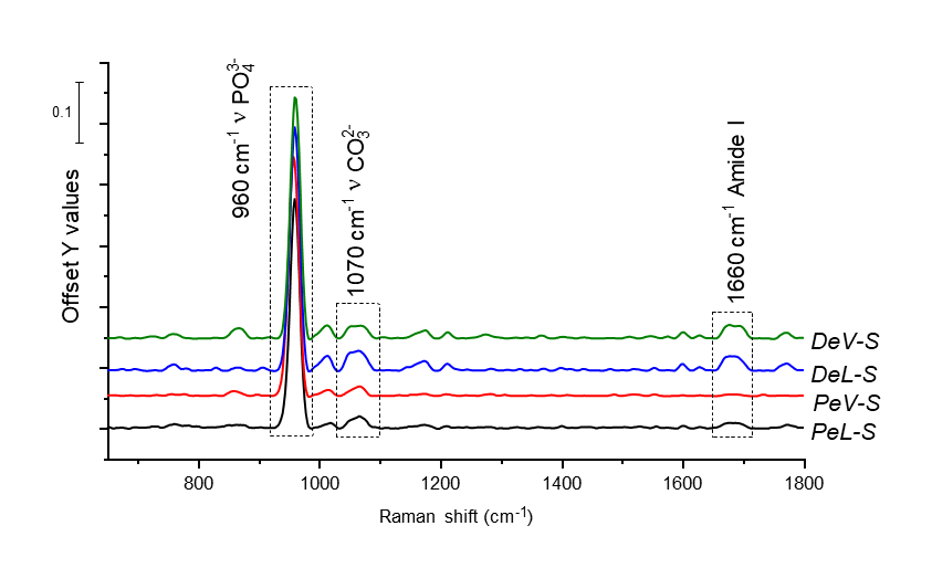
10.1016/j.saa.2021.119966
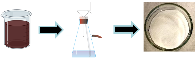
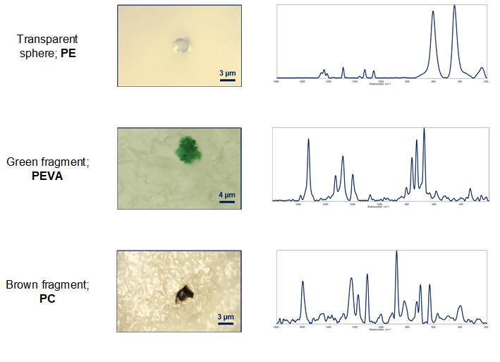
Filters inspection and Raman Microspectroscopy.
>2 mm Microplastics detection and characterization from human matrices
10.1016/j.envint.2020.106274
10.3390/polym14132700
10.3390/toxics11010040
10.1016/j.scitotenv.2023.165922
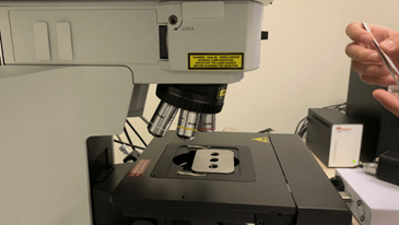
- Atomic Force Microscopy
-
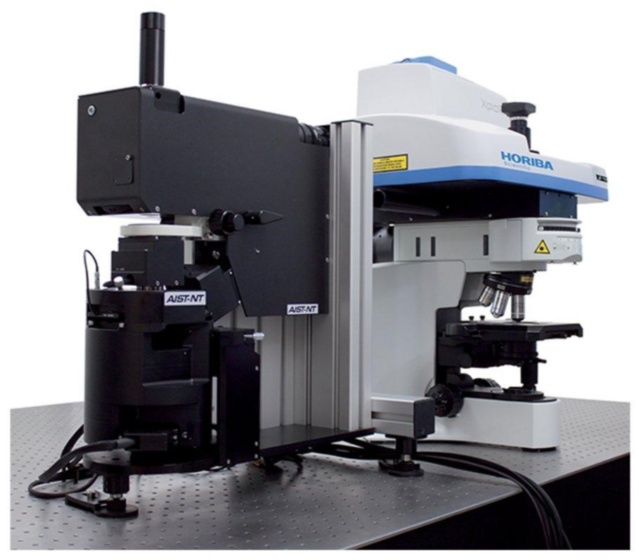
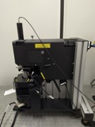
Atomic Force Microscopy (AIST-NT’s scanning probe microscopy SPM, Horiba Scientific)
 AFM is a powerful imaging technique that uses a sharp tip to scan a sample in order to obtain topographical images on the nanometer scale and quantifying local properties such as sample’s height and mechanical and electrical properties. The XploRA Nano platform integrates Atomic Force Microscopy together with the chemical information obtained from Raman spectroscopy.
AIST-NT’s SPM has a vertical resolution of less than nanometer (up to 0.1nm) and a lateral resolution of approximately (~ 30nm). The sample scanning range is 100 µm x 100 µm x 15 µm (±10 %) and the maximum sample size is 40 x 50 mm with 15 mm thickness. The range of the motorized sample positioning is 5 x 5 mm with a positioning resolution of 1 µm. The AFM platform allows fully-integrated use of confocal Raman microscopy and AFM for Tip-Enhanced Optical Spectroscopies such as Tip-Enhanced Raman Spectroscopy (TERS) and Tip-Enhanced PhotoLuminescence (TEPL), but also for co-localized AFM-Raman measurements. The myriad of AFM (Atomic Force Microscope) techniques that allow study of topographical, electrical and mechanical properties can be performed with any laser source available in Raman spectrometer. TERS and TEPL can provide nanoscale chemical and structural information, making the AFM-Raman platform a two-way road, where complimentary techniques provide novel and unique imaging capabilities to each other. 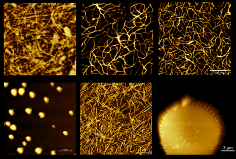
Top: Guanosine Hydrogel with different G:GMP ratio (Left 1:1, Center 1:2, Right 1:4).
Bottom left: Liposomes. Bottom center: Amyloyd fibrils. Bottom right: Diatom cell - Confocal microscopy
-
Confocal microscopy (confocal microscope A1, Nikon Instruments Inc, Melville, NY, USA,) Confocal microscopy permits to resolve detailed structures and morphology within tissues, cells or in whole mount preparations which are labeled with fluorescent probes. As main feature, confocal microscopy permits to acquire several optical sections within the specimens excluding out-of-focus signal and fluorescence background and thus increasing definition of the images and allowing high resolution 3D reconstruction. The instrument can be associated to different analytical techniques such as fluorescence in situ, FRAP and photobleaching to analyze proteins trafficking and protein targets. 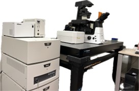
Confocal microscopy permits to resolve detailed structures and morphology within tissues, cells or in whole mount preparations which are labeled with fluorescent probes. As main feature, confocal microscopy permits to acquire several optical sections within the specimens excluding out-of-focus signal and fluorescence background and thus increasing definition of the images and allowing high resolution 3D reconstruction. The instrument can be associated to different analytical techniques such as fluorescence in situ, FRAP and photobleaching to analyze proteins trafficking and protein targets.
The A1R HD25 confocal microscope consist in assembly of 4 laser units (405 nm, 488 nm, 561 nm,640 nm), inverted microscope Ti-E2 and scanning head. It has a large field of view (25mm) and two different scanning system: Galvano and Resonant. Galvano mode increase quality of image and Resonant scanner increase imaging throughput and reduce phototoxicity of the sample for live imaging. The system is controlled through NIS-Elements C applications software able to process different acquiring images (Z-stack), suitable for photobleaching (FRAP), co-localization of multiple labeling (up to 4 channels) and photoactivation assay 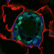
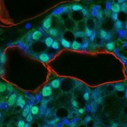
Triple staining of mouse mammary gland. In green a specific transcription factor was detected in the nuclei (blue) of epithelial cells. Fat pad of the gland is labelled in red. 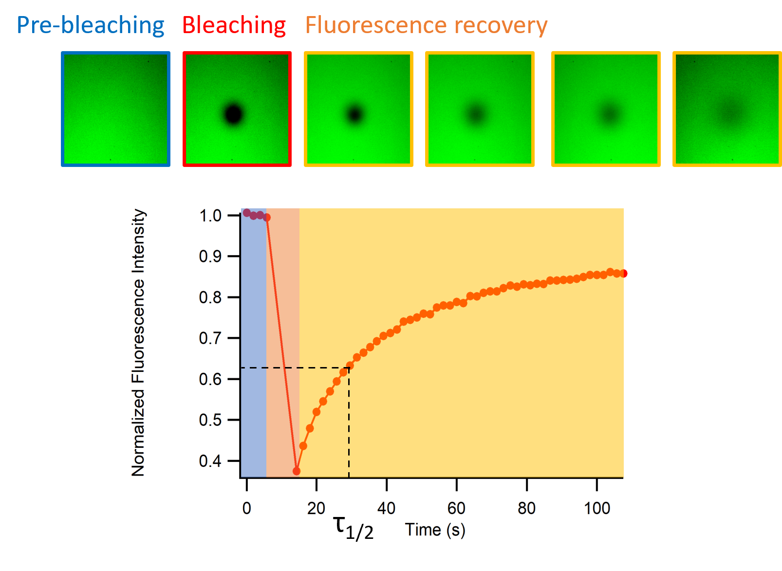
FRAP experiment on guanosine hydrogel 95% water content (G/GMP 1:1
- Automated microscope
-
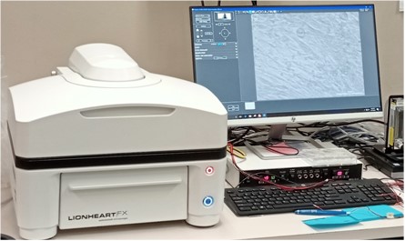
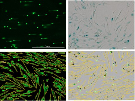
Automated microscope (Lionhearth FX Automated live cell Imager ,Biotek, BioTek Instruments, Inc., Vermont , USA)
The automated microscope is equipped with a set of lenses from 2.5x up to 100x magnification for whole organism to sub-cellular imaging. Furthermore, the instrument enables the assessment of kinetic live cell assay support with temperature and CO2/O2 control, humidity and reagent injectors. The instrument consists of a fully integrated microscope working in bright-field, phase contrast, color bright-field and fluorescence channels.
- Imaging Flow Cytometry
-
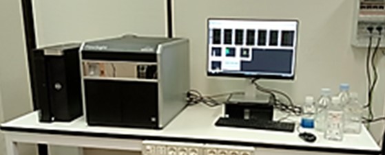
Imaging Flow Cytometry (Amnis Flow Sight, Luminex, Seattle USA)
Flow Cytometry is a potent technique able to obtain quantitative data in different biological samples. The fluidic system recreates a cell suspension stream where cells are excited with visible light or different lasers beams.
Thanks to cellular scattering and fluorescence it is possible to discriminate different cell populations and analyze specific cytosolic pathways. The imaging flow cytometer is an evolution of classic flow cytometry, in fact is equipped with a 20x magnification bright-field objective coupled with a CCD camera to acquire cell image in 12 channels to study cell morphology and fluorescence.
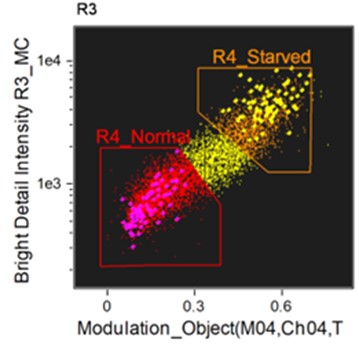
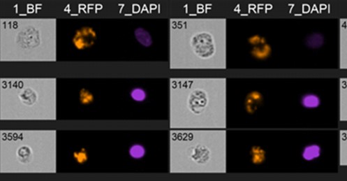
Flow sight resolution reach 1-2um. The instrument is equipped with 3 lasers (488 nm, 642 nm, 561 nm). The post- analysis software IDEAS allows the classification of digital images relative to each single acquired event utilizing 86 features per channels and 22 functions masks usable for analyze co-localization, nuclear translocation, apoptotic or mitotic DNA, cellular shape change etc. - Light Scattering Malvern Zetasizer Pro
-
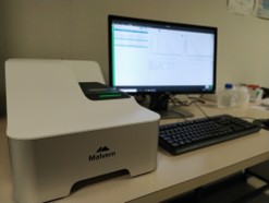
Light Scattering Malvern Zetasizer Pro
The Zetasizer Pro is a robust and versatile system for the measurement of particle and molecular size and electrophoretic mobility and zeta potential of nanoparticles. The MPT-3 Autotitrator accessory is designed to automate Zeta and/or Size versus pH titrations, helping to understand how your materials behave as a function of pH.
Minimum sample volume: 12 μl (DLS), 20 μl (Zeta Potential)
Minimum sample concentration: 0.1 mg/ml (NIBS)Minimum sample concentration: 10 mg/ml (forward angle)
Temperature control range: 0°C - 120°C (+/-0.5%)
Light Source: He-Ne laser 633nm, Max 5mW.
Laser safety: Class 1
Temperature: 15°C – 40°C
Humidity: 35% - 80% non-condensingSOURCE www.malvern.com
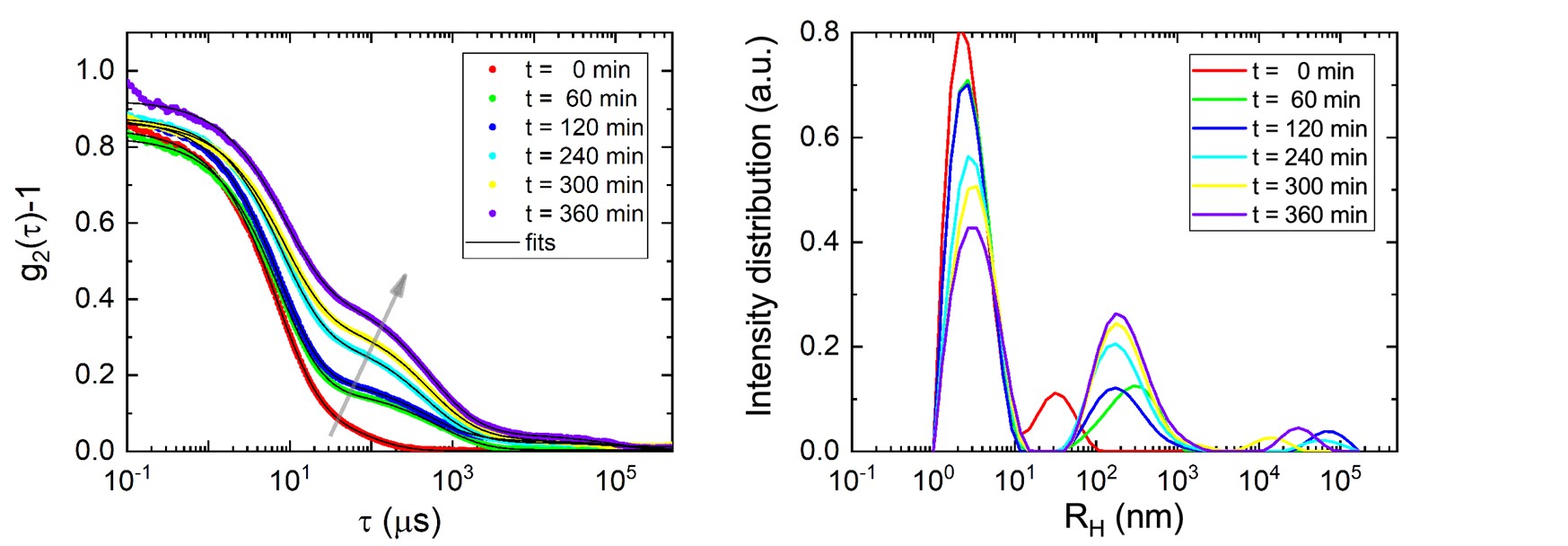
Left: Intensity Correlation functions of protein aggregation as a function of the time Right: Proteins Hydrodynamic radii as a function of the time
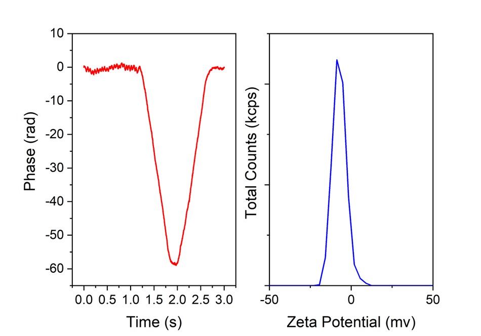
Left: Zeta Potential Phase diagram. Right: Zeta potential (mV